In our last post we gave you an insight about Human Respiratory System. In this post, I’d like to take you through the process of locomotion and movement.
Movement is change in posture or position. It is an essential and significant feature of living organisms. In many organisms’ different structures such as cilia, flagella, limbs etc show movement.
The movements which result in the change of place or location are called locomotion.
In this chapter we will study about the different types of locomotion and movement covering various aspects and processes involved in it.
All locomotions are movements but all movements are not locomotions.
MUSCLES
- It is specialized organ originated from mesoderm. The muscular tissues are made up of myocytes.
- A human body is made up of 639 muscles which have unique properties such as contractibility, excitability,elasticity and extensibility.
- They make almost 40-45% of body weight in adult human.
STRUCTURE
- A muscle sheet is covered with a sheath of connective tissue called epimysium.
- Inside perimysium, muscle has many muscle fibers arranged in bundles called fasciculi.
- Each fasciculi is covered by a sheath called perimysium.
- The muscle bundle are further bounded together by a common collagenous sheath of connective tissue called fascia.

TYPES OF MUSCLES

Difference between Skeletal, Smooth and Cardiac Muscle
| STRIATED OR SKELETAL MUSCLE | NON STRIATED OR SMOOTH MUSCLE | CARDIAC MUSLE |
| These are cylindrical. | These are spindle shaped. | These are cylindrical. |
| Ends are blunt. | Ends are tapering | Ends are blunt. |
| Fibres are unbranched. | Fibres are unbranched. | Fibred are branched. |
| Fibres occur in bundle. | Fibre occur singly. | They form 3-D network. |
| Blood supply in abundance. | Blood supply is poor | They are rich in blood supply. |
| Voluntary | Involuntary | Involuntary |
| Intercalated discs is absent. | Intercalated discs is absent. | Intercalated discs is present. |
| Innervated by branches from cranial and spinal nerves. | Innervated by autonomic nervous system. | Innervated by autonomic nervous system. |
| Multinucleated/ syncytial | uninucleated | uninucleated |
| Mitochondria are moderately abundant. | Mitochondria are fewer. | Mitochondria are abundant. |
| Myoglobin is abundant | Myoglobin ispoor. | Myoglobin is abundant |
| They contract quickly. | They contract slowly. | They show rhythmic contractions. |
| Easily get fatigued. | They do not fatigue. | They do not fatigue. |

STRIATED MUSCLES
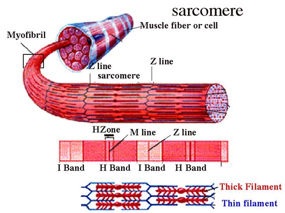
- Muscle fibre is covered by plasma membrane called sarcolemma, which encloses sarcoplasm which contains many nuclei.
- The muscle fibre contains parallel arranged myofibrils which have alternate light and dark bands.
- A single myofibril is made up of two types of myofilaments
- Thick myofilament– The thick myofilament consist of mainly myosin protein
- Thinmyofilament – thin myofilament consist of mainly actin protein.
- The endoplasmic reticulum present in sarcoplasm is called sarcoplasmicreticulum, which is store house of calcium ( required during muscle contraction)
- The dark band present on myofibril is called A band or anisotropic band
- The light band present on myofibril is called I band or isotropic band.
- At the center of dark A band, a comparatively lighter area called H band or hensen’s zone is present.
- A dark M line passes through the center of H band.
- The light I band consist of dark line which passes through the center called Z- line.
- The part of myofibril between two successive Z line is called sarcomere.
The sarcomere is the structural and functional unit of myofibril. A sarcomere comprises of a single A band and half of each adjacent I- band.
STRUCTURE OF CONTRACTILE PROTEIN
THICK MYOFILAMENT
- The thick myofilaments are made up of polymerised protein called myosin. The monomeric proteins called meromyosins polymerize to form myosin protein.
- Each meromyosin have two parts
- A globular head with a short arm
- A tail.
- The head along with short arm called heavy meromyosin(HMM) , it has a site for binding of actin and ATP. It acts as an ATPase enzyme . It hydrolyses ATP to produces energy.
- Tail is called light meromyosin(LMM).
THIN MYOFILAMENT
These are made up of 3 proteins—actin, tropomyosin and troponin.
- ACTIN
- It is globular protein.
- It has low molecular weight.
- It occurs in two forms- monomeric g- actin and polymeric f-actin.
- The g- actin polymerises to form f actin in presence of magnesium ion.
- TROPOMYOSIN
- It is fibrous molecule.
- The two filaments run closely along each other throughout entire length.
- At resting state it separates the actin and myosin by binding to myosin binding site on actin filament and hence prevent formation of cross bridges which in turn prevent contraction of muscle fibre.
- TROPONIN
- At regular intervals of tropomyosin a complex protein called troponin is present. It masks the active binding site for myosin on actin filament.
- It is trimeric protein( it has 3 units which acts as different components)
TroponinI: it inhibits actin- myosin interaction and bind to other components of troponin.
Troponin T: it is binding site for tropomyosin.
troponin C: it is binding site for calcium.
MECHANISM
- The mechanism of muscle contraction was explained by a theory called sliding filament theory. According to this theory, the contraction of muscle fibre occurs when thin filament i.e. actin filament slides over thick filament i.e.The myosin filament.
- The contraction of muscle is initiated by the signal sent by CNS via motor neuron. When impulse reaches the axon terminal .The vesicles containing neurotransmitters fuses with axon membrane.After fusion they release neurotransmitter, acetylcholine; which travels through synaptic cleft and generate action potential in sarcolemma.
- The impulse generated then spread from sarcolemma to the T- tubules. The impulse stimulates sarcoplasmic reticulum to release action potential in the sarcolemma.
- An increase in Ca2+ concentration in sarcoplasm starts the filament sliding( if concentration decrease the sliding turns off ). When a muscle fibre is relaxed (not contracting ), the concentration of Ca2+ in sarcoplasm is low. This is because
- Sarcoplasmic reticulum membrane contains Ca2+ active transport pumps that move Ca2+ from sarcoplasm to sarcoplasmic reticulum.
- As muscle action potential travels along the sarcolemma, Ca2+ channels open in the SR membrane. As a result Ca2+ floods into sarcoplasm around thick and thin filaments. The
- Ca2+ released from sarcoplasmic reticulum combine with troponin, causing it to change shape.
- The shape moves the troponin- tropomyosin complex away from myosin- binding sites.
- The globular head of myosin acts as an ATPase enzyme and hydrolyses ATP. The energy derived from the hydrolysis of ATP is used by myosin to bind the exposed active site on actin filament to form a cross bridge.
- This pulls actin filament towards the centre of A band. The Z line also pulled inwards causing shortening of sarcomere. The thin myofilaments move past the thick myofilaments due to which H zone narrows. This reduces the length of I band but retains length of A band. Again the ATP binds to the myosin and cross bridge between actin and myosin is broken.
- The ATP is again hydrolysed and the cycle of formation and breakage of cross bridges is repeated causing sliding. The process continues till the calcium ion is pumped back into the sarcoplasmic reticulum.

MUSCLE RELAXATION
After contraction of muscle the calcium moves back into sarcoplasmic reticulum. The amount of calcium is reduces in sarcoplasm due to which calcium does not bind with troponin C. The troponin undergoes change in shape and tropomyosin and troponin attain their earlier position and state. This blocks the active site of myosin on actin and myosin does not bind to actin. This causes relaxation of muscle.
ALL OR NONE PRINCIPLE
A minimal strength of a stimulus required to cause the contraction of a muscle fibre brings about maximum contraction and no further increase in contraction would occur by increasing the strength of the stimulus.
SINGLE MUSCLE TWITCH
A single quick isolated contraction of a muscle fibre to a single stimulus of threshhold value is called single muscle twitch.
MUSCLE FATIGUE
The repeated contraction of skeletal muscle anaerobically leads to accumulation of lactic acid in muscle. The lactic acid accumulated in myocytes then diffuses in to the blood and transport to liver. In liver 4/5th of lactic acid is converted to glycogen by the process gluconeogenesis and 1/5th lactic acid is oxidized to co2 and water.
RIGOR MORTIS
Extreme rigidity of body after death is called Rigor mortis. It is due to complete depletion of ATP and phosphocreatine.
Important : The phenomenon of rigor mortis was first described in 1811 by the French physician, P.H. Nysten, but its physiological basis was not discovered until 1945 by Szent-Györgyi .
Take this Quiz on Locomotion and Movement and see how you fare!! All the best 🙂
In our next post we will discuss Skeletal System in detail and also some of the disorders associated with it. Happy Reading 🙂
Leave a Reply