An adult human being has 206 bones and this number is more in case of a child as there are a number of unfused bones including the bones of sacrum and coccyx of the vertebral column. The skeletal system of humans can be classified into two categories- the axial skeletal system and the appendicular skeletal system. Before studying them in detail, let us first study various types of bones present in the body:
1-Long bones: The bones which are much longer in length than their width are termed as long bones. Example-femur, humerus, radius, ulna etc.
2-Short bones: Short bones have almost similar size in terms of length and width. Example-carpals and tarsals etc.
3-Flat bones: These are more of a sheet like shape having spongy bone tissue sandwiched between two layers of compact tissue on dorsal and ventral side. Example-cranial bones, scapula and sternum etc.
4-Irregular bones: The bones which do not have a definite shape are classified as irregular bones. Example-mandible.
5-Sesamoid bones: Sesamoid bones appear in tendons as a result of friction and tension. They do not have a definite shape and size and are mostly found at the joints. Their number varies from person to person. Example-patella
Axial skeleton
The axial skeleton lies in the main axis of the body excluding the appendages. There are a total of 80 bones in the adult axial skeleton and they can be distributed as Skull (8 cranial and 14 facial bones), hyoid bone, ear ossicles (6), vertebral column (26 if fused and 33 if unfused), thorax (24 ribs and a sternum).
Skull
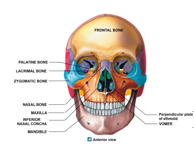
Skull contains 22 bones excluding the ear ossicles. There are 8 bones that constitute the cranium (cranial bones) and 14 facial bones. The cranial bones include- a frontal bone, 2 temporal bones, 2 parietal bones, an occipital bone, sphenoid and the ethmoid bone. The bones of face include- a mandible, 2 maxillae, 2 zygomatic bones, 2 palatine bones, 2 nasal bone, 2 lacrymal, 2 inferior nasal conchae and a vomer bone.
Facial bones:
1-Mandible: Mandible is the bone of lower jaw. It consists of a curved body which has two nearly perpendicular structures called the ramus. The ramus has a condylar process on the posterior side, a coronoid process on the anterior side, and a u-shaped depression between the two processes called the mandibular notch.
2-Maxillae: Maxilla is the bone of upper jaw and is paired. It articulates with every bone of the face except mandible. It has an alveolar arch having alveoli or sockets into which teeth fit. The fusion of two maxillae occurs before birth and if it fails the condition is known as cleft palate.
3-Zygomas: Zygomas or zygomatic bones are the paired bones also known as cheek bones. It has a posterior projection called temporal process of zygomatic bone which articulates with the zygomatic process of temporal bone.
4-Nasal bones: These are also paired bones, rectangular in shape that form the nasal bridge.
5-Lacrimal bones: These are paired bones located posterio-laterally to the nasal bone. They are the smallest bones of the face.
6-Palatine bones: These are paired L-shaped bones whose horizontal plates form posterior portion of the hard palate.
7-Inferior nasal conchae: These are two bones located inferior to the middle nasal conchae of the ethmoid bone. They do not form a part of the ethmoid bone.
8-Vomer: Vomer is an unpaired bone which forms the inferior portion of the nasal septum. On its superior end, it articulates with the perpendicular plate of the ethmoid and sphenoid bone and inferiorly with maxillae and palatine bones.
Bones of cranium:
1-Frontal bone: Frontal bone forms the forehead and also the roof of eye orbits. It gradually slopes down, straightens and thickens forming the supra-orbital ridges above the orbits.
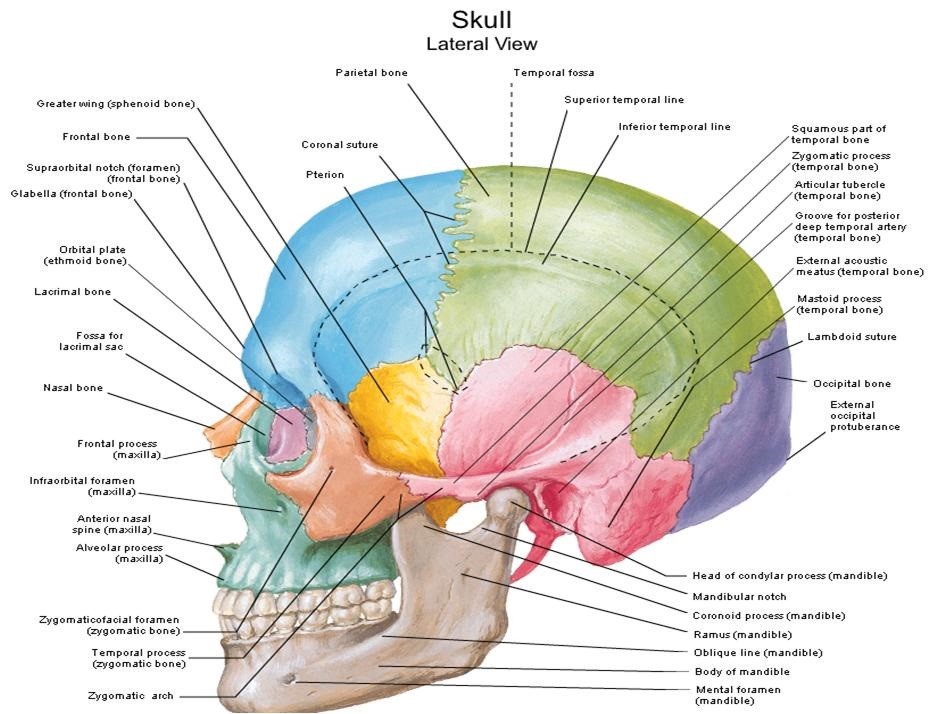
2-Parietal bones: There are two parietal bones that form the roof of cranium and also extend towards the sides.
3-Temporal bones: These are two bones located on either side of the skull above the ear, the area commonly referred to as temple. The inferior portion of the temporal bone projects anteriorly and forms the zygomatic process of temporal bone.
Important structures of temporal bone:
Zygomatic arch: Formed by articulation of zygomatic process of temporal bone and temporal process of zygomatic bone.
Mandibular fossa: A notch in zygomatic process of temporal bone, posterior to articular tubercle, that receives condylar process of mandible.
External auditory meatus: The ear canal located posterior to temporo-mandibular joint.
Mastoid process: Rounded protuberance of temporal bone located posterior and inferior to external auditory meatus.
Styloid process: It is a pointed projection that extends inferiorly from the inferior region of temporal bone below the external auditory meatus.
4-Occipital bone: It is located on the posterior region of the skull and constitutes to a major portion of the skull.
Picture Reference : Source
Picture Reference : Source
Important structures of occipital bone include:
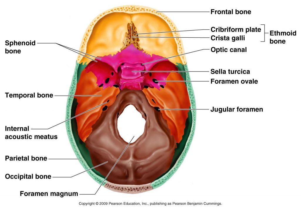
Foramen magnum: It is the largest hole of human body that allows articulation of brain and spinal cord. It is located on the posterior portion of the skull, in the inferior region of occipital bone.
Occipital condyles: These are two oval structures located on the either side of foramen magnum, they facilitate articulation of skull with the facets present on atlas, first vertebrae.
Picture Reference : Source
External occipital protuberance: It is a bony protuberance located in the occipital bone just above the foramen magnum. It can be felt as a large bump on the back side of the neck just above the head.
Nuchal lines: These are a pair of two bony ridges that extend laterally from the external occipital protuberance. The upper pair is the superior nuchal lines and the lower one is called inferior nucha lines. They provide site for attachment of muscles.
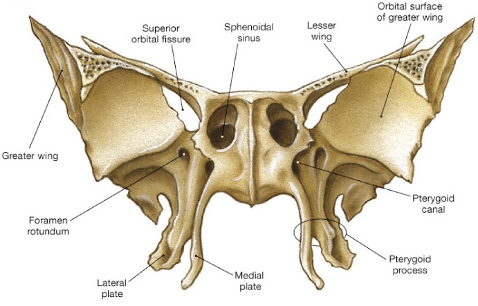
5-Sphenoid bone: It is a butterfly shaped bone located on the midregion of the base of the skull. Sphenoid forms an important part of the floor of cranium as it articulates with all other bones and holds them together. It has a saddle-shaped body at the center, two large great wings extending laterally, two smaller lesser wings, two pterygoid process which project inferiorly from the point of articulation of greater wings and the body.
Picture Reference : Source
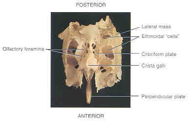
6-Ethmoid bone: It is located anteriorly to sphenoid and forms a part of cranial floor as well as a major portion of nasal cavity.
Picture Reference : Source
Important structures of ethmoid include:
Cribriform plate: It lies in the anterior part of the cranial floor and forms the floor of nasal cavity.
Crista galli: Triangular process that projects superiorly from the cribriform plate.
Perpendicular plate: It extends from the inferior portion of cribriform plate and forms the superior portion of the nasal septum.
Lateral masses: Form the major portion of the wall between the nasal cavity and orbits. They have two thin, scroll-shaped projections that extend laterally to the nasal septum; superior nasal concha and middle nasal concha.
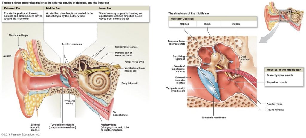
Ear ossicles
There are three small bones present in the mid-region of each ear:
1-Malleus: It is also known as hammer. The handle of malleus articulates with the internal surface of tympanum (ear drum). The head of malleus articulates with the body of incus.
2-Incus: Also known as anvil, incus is the middle bone that articulates with the head of stapes.
3-Stapes: Also known as stirrup, it is the innermost bone of the ear which fits into the oval window. Stapes is the smallest bone in the human body.
Picture Reference : Source
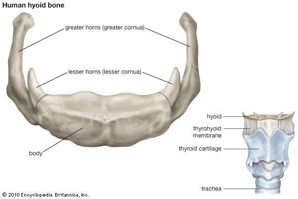
Hyoid bone
Hyoid is the only bone of the body that does not articulate with any other bone. It is located between the mandible and larynx. It is U-shaped bone consisting of a body in the anterior side, two small paired lesser horns posterior to the superior hyoid and two posteriorly extending greater horns. Hyoid bone is easily fractured in case of strangulation.
Picture Reference : Source
Read part 2 of this post to know about vertebral column in depth. In case of any doubt please feel free to drop in your query in the comment box below, we will get back with an answer in a jiffy. Happy Reading 🙂
Leave a Reply