Pelvic Girdle
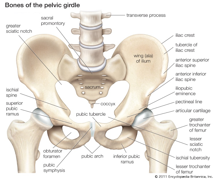
Pelvic girdle is formed by two bones i.e. coxal bones or os coxa. The two coxal bones are joined anteriorly by a cartilagenous tissue called pubic symphysis. Posteriorly the coxal bones articulate with the auricular surface of sacrum to form sacro-illiac joint. The pelvic girdle along with the sacrum forms the pelvis. Pelvic girdle provides site for articulation of femur of hindlimbs and in this way connects lower limb to axial skeleton.
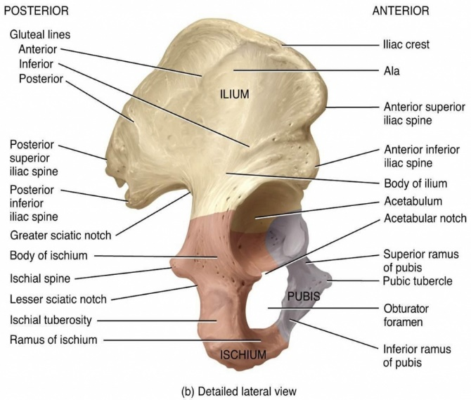
Each coxal bone is formed of 3 bones-Ilium, Ischium, and Pubis. These bones initially exist as three separate bones and fuse by the age of 23.
Picture Reference : Source
Ilium
Ilium is the largest of the coxal bones. It has a superior wing called ala and an inferior body. The superior border of illium is known as the iliac crest which ends anteriorly, on the lateral side, as anterior superior iliac spine. A shallow depression inwards, and below lies the anterior inferior iliac spine. Posteriorly, the iliac crest terminates into a bony posterior superior iliac spine, below which is the posterior inferior iliac spine. Below the posterior inferior iliac spine lies the greater sciatic notch through which the sciatic nerve passes. The medial surface of ilium has an iliac fossa, posterior to which lies the iliac tuberosity. Inferior to the iliac tuberosity, is the auricular surface of ilium via which it articulates with sacrum. Anteriorly and inferior to auricular surface is a prominent ridge margin called arcuate line.
Ischium
Ischium contributes the inferior portion of the hip bone posteriorly. It has a superior body and an inferior ramus. Its body articulates with the body of ilium and ramus fuses with pubis. Ischium has a projecting ischial spine inferior to greater sciatic notch, below which is the lesser sciatic notch. The inferior most portion of the ischium is called the ischial tuberosity, which can be felt when a person sits, the reason why ischium is called ‘the sit bone’. The ached ramus and pubis fuse to form the obturator foramen, the largest foramen in the skeleton.
Pubis
Pubis forms the anterior-inferior portion of the coxal bone. It has a superior ramus, a body, and an inferior ramus. The superior border of pubis, anteriorly, is called the pubic crest. On the lateral side of the pubic crest is the pubic tubercle which marks the beginning of a ridge called pectineal line, which projects superiorly and fuses with arcuate lines of the ilium. The two pubic bones are joined at pubic symphysis, inferior to which is the inferior ramus of pubis. Together, portions of ilium, ischium and pubis fuse to form a round cavity called acetabulum, on the lateral side of pelvis. Inferior to acetabulum, is the acetabular notch at which ligaments of femur articulate with pelvis.
Hindlimb
The two hind limbs have 60 bones, each having 30. The bones of hindlimb include-Femur, Patella, Tibia and Fibula, 7 tarsals, 5 metatarsals and 14 phalanges.
Femur
Femur is the longest and the strongest bone of body. It is present in the proximal part of hindlimb. Proximally, it articulates with acetabulum of pelvic girdle via its head, and distally with tibia. Femur has a long shaft inclined towards the mid-sagittal plane.
1-Anterior view: The proximal end of femur has a round ball-like protuberance called the head. The head of femur has a small cavity on its surface called fovea capitis. Laterally, the head constricts into neck and lateral and inferior to the neck is a projection called the greater trochanter. The neck continues into shaft and medial and inferior to the shaft is the lesser trochanter. The diagonal line joining the two trochanters is called inter-trochanteric line. The shaft is smooth on the anterior surface. The distal end of femur has two protuberances called the lateral and medial condyles which articulate with the respective condyles of tibia. Superior to the two condyles of femur are the lateral and medial epicondyles on the respective sides. Superior to medial epicondyle is the adductor tubercle to which adductor magnus muscle attaches. Between the lateral and medial condyle is the intercondylar sulcus. The anterior surface between the condyles is called the patellar surface.
2-Posterior view: Posteriorly, the shaft of femur is rough and highly rugged. A broad ridge in the area between the two trochanters is called intertrochanteric crest. Inferior to this crest, laterally, is a long ridge running down the shaft called the gluteal tuberosity. Medial to gluteal tuberosity is another vertical ridge called linea aspera. On the posterior side, there is a depression in the area between the two condyles, the intercondylar fossa.

Picture Reference : Source
Patella
Patella is a sesamoid bone present anterior to the point of articulation of tibia and femur. It is also known as knee-cap. It has a broad proximal end called the base and a sharp distal end called the apex. The posterior surface of patella has two articular facets present on the lateral and medial sides for articulation with the respective condyles of femur. It also articulates with tibial tuberosity via ligaments.

Picture Reference : Source
Tibia
Tibia is the bone of lower hindlimb. It is also known as shin bone. Tibia articulates with femur and fibula at its proximal end and with talus bone of the ankle and fibula at its distal end.
The proximal end of tibia has two concave condyles called the lateral and medial condyles which articulate with the respective condyles of femur. There is an upward prominence between the two condyles called intercondylar eminence. Inferior to the condyles is a roughened triangular area called tibial tuberosity. Fibula articulates with tibia at inferior region of the lateral condyle. The shaft of tibia has a sharp anterior border which can be felt in the shin region. The distal end of tibia has a sharp projection on the medial side called the medial malleolus. It articulates with talus bone of the ankle. On the lateral side of the distal end is a facet for fibular articulation called the fibular notch. The base of tibia at the distal end is faceted for articulation with talus.
Fibula
Fibula is the second bone of lower hindlimb. It is located laterally but slight backwards and parallel to tibia. It does not directly articulate with femur. The head of fibula, expanded in all directions, articulates with inferior part of lateral condyle. It has a styloid process pointing upwards and posterior. The head is constricted in the inferior region called the neck. Fibula has a slender shaft with multiple borders. The distal end of fibula has a sharp projection on the lateral side called the lateral malleolus. Just above the distal end is a triangular area, whose apex when traced upwards continues into anterior border of fibula. Medial to the anterior border is the medial border. The area between the anterior and medial border is called medial surface. On the posterior side is the posterior border. The area between the anterior and posterior border is called the lateral surface. Posterior surface is the largest surface and lies between the medial and posterior border. At the distal end, inferior to medial surface, anteriorly, is a triangular facet for articulation with talus. Posterior to this is the malleolar fossa.

Picture Reference : Source
Tarsals
There are 7 tarsal bones. The proximal region of tarsals, the tarsus, is situated posteriorly. This region has two bones-talus and calcaneus. Calcaneus is the strongest tarsal bone, palpable in the heel-region.
The anterior tarsal bones include-navicular, lateral cuneiform, intermediate cuneiform, and medial cuneiform, and a cuboid bone located lateral to lateral cuneiform.

Picture Reference : Source
Metatarsals
The sole region of foot has five metatarsal bones numbered 1-5 in medial to lateral order. Each metatarsal has a proximal base, intermediate shaft, and a distal head. Metacarpals articulate proximally with three cuneiform bones and cuboid bone and distally with first row of phalanges.
Phalanges
There are a total of 14 phalanges that form bones of toes of the foot. Each bone is referred to as phalanx. the great toe has two phalanx while the others have 3 phalanx each.
References
1-Principles of anatomy and physiology, Gerard Tortora and Bryan Derrickson, International student version.
Leave a Reply