Living cells require constant supply of nutrients O2 and other essential substances. Also, waste or harmful substances produced; need to be removed continuously for healthy functioning of tissues. It is therefore, essential to have efficient mechanism for the movement of these substances to the cells and from the cells. Human beings have blood which is most commonly used body fluid for this purpose. Lymph is another body fluid which helps in transport of certain substances. In this article, we will study about human circulatory system where in we will talk about blood composition, properties of blood and mechanism of blood circulation.
BLOOD
It is the connective tissue consisting two parts:
- PLASMA– the fluid matrix; constitutes 55%
- FORMED ELEMENTS– the cellular part; constitutes 45%

PLASMA: It is a straw colored, viscous fluid forming matrix of the blood.
Composition:
1.Water- 90-92%
2.Proteins- 6-8%
- Fibrinogen- it is an important clotting factor.
- Globulins- they are involved in defense mechanism and also produce immune responses.
- Albumins- they help in osmotic balance. They maintain blood volume and pressure.
3.Minerals: plasma contains small amounts of minerals like Na+, Ca++, Mg++, Cl– etc.
4. Glucose, amino acids, lipids etc. are also present in plasma as they always transit in the body.
Plasma without clotting factor is called as serum.

FORMED ELEMENTS
These include erythrocytes, leucocytes and thrombocytes.
Erythrocytes [ RBCs – RED BLOOD CELL]:
- Most abundant
- Number- 5 million to 5.5 million of RBCs mm-3
- Biconcave and anucleated.
- Consist red pigment called ‘haemoglobin’
- Quantity of haemoglobin: 12-16 g in every 100ml of blood
- The process of formation is called erythropoiesis
LEUCOCYTES [ WBCS- WHITE BLOOD CELLS ]:
- Number: 6000-8000 mm-3
- Have nucleus.
- Leucocytes containing granules in their cytoplasm called granulocytes and those which are clearly visible are called agranulocytes.
- Formation process is called leucopoiesis.
AGRANULOCYTES:
LYMPHOCYTES:
- 20-25% of total WBCs
- 2nd most numerous type of leucocytes.
- They exist in two major forms named B and T lymphocytes which are responsible for immune responses.
MONOCYTES:
- Largest leucocytes.
- They mature and become macrophages which are phagocytic (that engulf bacteria and other cellular debris)
GRANULOCYTES:
EOSINPHILS
- 2-3% of WBCs
- Have antihistaminic properties.
- Resist infections and associated with allergic reactions.
BASOPHILS:
- Least abundant (0.5 – 1 %)
- Secretes serotonin, heparin(anti-coagulant) and histamine(vasoconstrictor)
NEUTROPHILS:
- Most abundant cells (60-65%)
- Phagocytic
Thrombocytes [platelets]:
- Number- 1,50,000-3,50,000 mm-3
- cell fragments rather than true cells.
- Formed in bone marrow and called as megakaryocytes.
- Plays important role in blood clotting.
MECHANISM OF BLOOD COAGULATION
- An injury stimulates platelets in blood to release coagulation promoting substance called thromboplastins which activates mechanism of coagulation.
- Thromboplastins help in formation of enzyme complex thrombokinase.
- Thrombokinaseconverts inactive protein into prothrombin and then into thrombin.

- Thrombin is an enzyme which converts fibrinogen into fibrin.

- Fibrin forms network of threads which traps dead and damaged formed elements to form blood clot.
- The clot seals the wound in the vessel to stop bleeding. This is called blood clotting.
LYMPH
- The fluid is collected and drained back to the major veins by an elaborate network of vessels called lymphatic system. The fluid present in lymphatic system is called lymph.
- Lymph is the colorless fluid containing specialized lymphocytes responsible for immune responses.
- Lymph is filled through lymph nodes which are abundant in neck, groin and armpits.
- Lymph nodes consists
- phagocytic cells which removes pathogens. Tonsils, thymus and spleen are called as lymph nodes. They are called as lymphoid organs.
FUNCTION:
- It transports oxygen, nutrients; hormones etc. to the body parts and bring back carbon dioxide and other waste products.
- They destroy the invading microorganisms.
HUMAN CIRCULATORY SYSTEM
It is called as blood vascular system which consists of a muscular chambered heart, a network of closed branching blood vessels and blood.
HEART:
- It is located in thoracic cavity, slightly tilted to left.
- It is protected by double membranous wall called pericardium. A space is called pericardial cavity which is present between two layers which is filled with fluid called pericardial fluid.
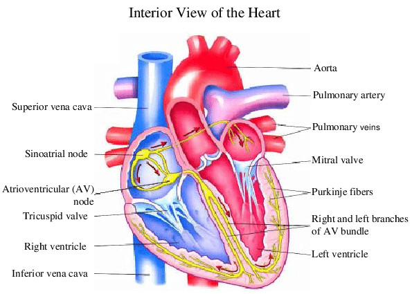
- Heart is divided into 4 chambers. The smaller chambers called atria and other two lower chambers called ventricles. The walls of ventricles are thicker than that of atria.
- A thick fibrous tissue called antrio ventricular septum separates the atria and ventricle of the same sides. Atria are separated by interatrial septum and ventricles by inteventricular septum.
- The opening between atria and ventricles are guarded by atrioventricular valves. The AV valve between right atrium and ventricle has 3 flaps therefore called tricuspid valve. The AV valve between left atrium and ventricle has two flaps and therefore called bicuspid valve or mitral valve.
- Three semilunar valves are found at the points where pulmonary artery(arising from right ventricle and carrying deoxygenated blood to the lungs) and aorta(large artery arising from left ventricle are carrying oxygenated blood to all parts of the body) leave the heart. These valves prevents backflow of the blood.
- The right atrium receives deoxygenated through coronary sinus and two large veins called venae cavae( one superior and one inferior). The left atrium receives oxygenated blood from the lungs through two pairs of pulmonary veins.
- Our heart beat normally 70-75 times in a minute which is called heart rate.
One cardiac cycle is of 0.8 seconds.
- During cardiac cycle each ventricle pumps out approximately 70 ml of blood. This is called stroke volume. The stroke volume is multiplied by the number of beats giving cardiac output which is 72×70=5040mlper minute i.e. about 5 L per minute.
DOUBLE CIRCULATION:
This refers to that blood passes through heart twice for each cycle. This includes systemic and pulmonary circulation.
Pulmonary circulation: The deoxygenated blood pumped into pulmonary artery is passed onto the lungs from where oxygenated blood is carried by the pulmonary veins into the left atrium.
Systemic circulation: the oxygenated blood entering the aorta is carried by network of arteries, arterioles and capillaries to tissue from where deoxygenated blood is collected by network of veins, venules and vana cava which is emptied into right atrium. Thus, systemic circulation provides nutrienrts, oxygen and other essential substances to the tissues and take CO2 and other harmful substances away for elimination.

| ARTERIES | VEINS | CAPILLARIES |
| They distribute blood from heart to various parts. The walls are thick and muscular. They have no valves. The flow is fast as blood in them is under great pressure. They carry oxygenated blood except pulmonary artery | They collect blood from different parts and pour into the heart. They have thin walls. They have valves. The flow is not so fast and blood is under low pressure. They carry deoxygenated blood except pulmonary vein | They are narrowest blood vessels, through which exchange of gases and nutrients between blood and tissue occurs |
CONDUCTING SYSTEM IN HEART
- The entire heart is made up of cardiac muscles. A specialized cardiac musculature called as nodal tissue which is distributed in heart.
- A patch of this tissue called sino-atrial node(SAN) present in the upper right corner of the right atrium. Another named atrio-ventricular node(AVN) present in lower left corner of right atrium.
- A bundle of nodal tissue i.e. atrio ventricular bundle (AV bundle) divides into right and left bundle. These branches give rise to minute fibrespurkinjefibres. These fibresalongwith right and left bundle known as bundle of his.
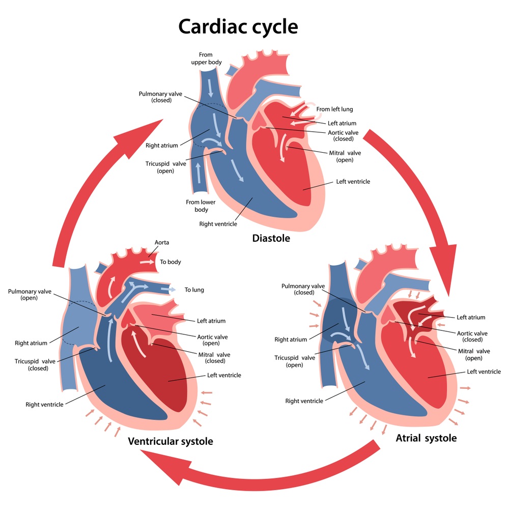
- The cardiac cycle refers to repeating pattern of contraction and relaxation of heart. The phase of contraction is called as systole and the phase of relaxation is called as diastole.
- It begins with joint diastole when all four chambers are at rest. Then atrial systole takes place, this results in increase of blood flow into the ventricles. Then ventricular systole occurs which coincides with atrial systole again. Then the blood flows from ventricles to aorta and pulmonary artery. Now, ventricular diastole takes place.
- The sequential events cyclically repeated in the heart constitute cardiac cycle.
- During cardiac cycle two prominent sounds are produces which can be heard by stethoscope named as lubb and dubb.
DISORDERS
- Hypertension [high blood pressure]: a blood pressure of 120/80 is considered as normal. If the blood pressure is 140/90 or higher, it shows hypertension and it affects vital organs brain and kidney.
- Angina pectoris: a symptom of acute chest pain when no enough oxygen is reaching the heart muscle.
- Heart failure: state of heart when it does not pump blood effectively enough to meet the needs of the body. It is also called as congestive heart failure because congestion of lungs is main symptom of the disease.
- Heart attack: it occurs when heart muscles are suddenly damaged by inadequate blood supply.
- Cardiac arrest: it means complete stoppage of heart beat.
READ OUR NEXT POST – HUMAN RESPIRATORY SYSTEM
Leave a Reply